ESB Scientific Image Competition 2022
The judges would like to thank everyone who took part in ESB-SIC22, the entries were all of a high quality, and reflect the range of scientific work being undertaken by our younger members. All of the entries are shown below the winners – please click on images for credits, and a precis of the science they depict!
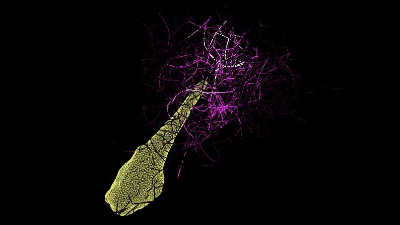
Winner: Rémy Gauthier
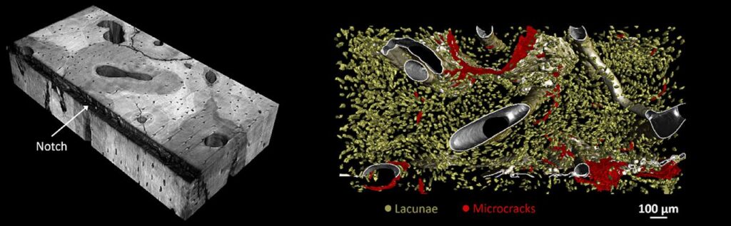
Human cortical bone crack propagation is influenced by bone microarchitecture by Rémy Gauthier, Université Gustave Eiffel – Université Claude Bernard Lyon 1.
This figure is a 3D rendering of a human radius cortical bone samples that, scanned with synchrotron radiation micro-computed tomography at the ESRF (voxel size = 0.7 µm). Previously to the image acquisition, this bone sample was subjected to a toughness test under quasi-static loading in order to quantify the energy needed to propagate a crack. The observed features are the vascular network, the osteocyte lacunae (yellow) and the cracks (red). The high radius toughness was associated with its capacity to deviate cracks through its complex microarchitecture, as illustrated in the image where the crack follows the cement line.
Publication link: https://doi.org/10.1016/j.jbiomech.2019.01.008
Second place: Graciosa Teixeira
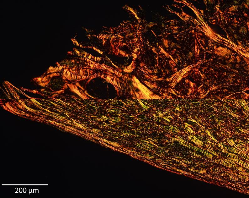
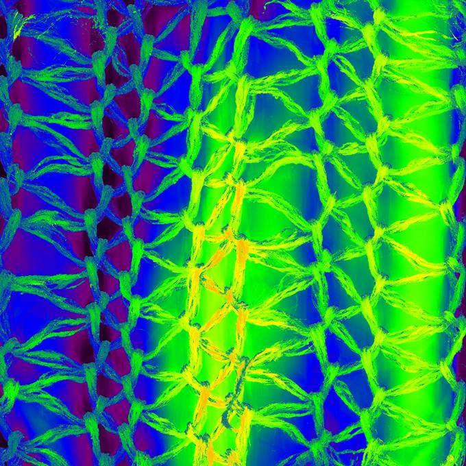
Meniscus ageing – severely and mildly degenerated collagen in human meniscus by Graciosa Teixeira, Institute of Orthopaedic Research and Biomechanics, Ulm University, Ulm, Germany.
Representative picrosirius red staining for collagen content in severely (upper image) versus mildly (lower image) degenerated human menisci imaged under polarised light. Changes in collagen composition with ageing/degeneration lead to a mechanically incompetent tissue under compression. The severely degenerated meniscus is from a 70-year-old patient depicting strong signs of collagen loss and disorganization, whereas the mild degenerated sample corresponds to a 24-year-old injury patient with little collagen degradation. Mature collagen fibres appear red/yellow, being mostly associated with collagen type I. Thin/younger fibres appear green and are generally associated with collagen type III (scale bar, 200 µm).
Third place: Gustavo Orozco
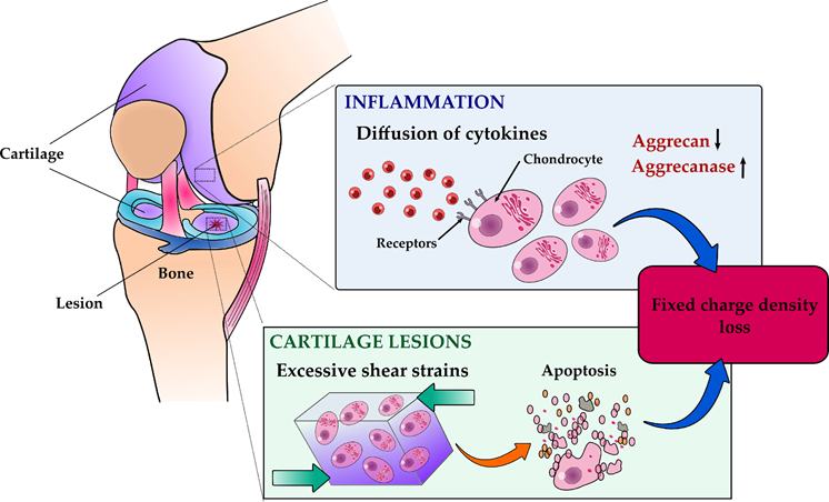
Cartilage degeneration mechanisms after ACL reconstruction by Gustavo Orozco, Lund University.
Anterior cruciate ligament injury can lead to post-traumatic osteoarthritis. One sign of PTOA is proteoglycan loss (PG) of cartilage. Despite ACL reconstruction, altered biomechanics and prolonged joint inflammation, and associated diffusion of cytokines (such as interleukins (ILs)) could alter the balance between anabolic and catabolic activities of cartilage and decrease the amount of PG. Eventually, this may speed up tissue degeneration and decrease the fixed charge density of PGs. Hence, we have developed a numerical degeneration model using a 3D patient-specific ACL reconstructed knee to estimate FCD decrease via diffusion of pro-inflammatory IL cytokines.
Highly commended
Above: A gallery of images submitted by ESB members to the ESB Scientific Image Competition, 2022. Click on any image for a larger/complete version and more details.